Microscopes BRESSER
All Microscopes Advanced filters → |
Popular modelsCompare using chart →
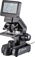
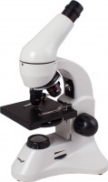
training, laboratory, biological, optical-digital, increase 30 – 1200 x, lens 4x, 10x, 40x(s), eyepiece 7.5x, movable table, camera, connection: HDMI, USB, card reader
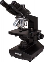
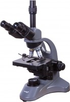
laboratory, biological, optic, increase 40 – 2000 x, lens 4x, 10x, 40x(s), 100x(s) oil, eyepiece paired PLAN WF10x, PLAN WF20x, swivel eyepiece, keller lighting, movable table
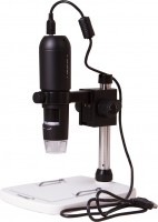
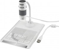
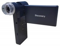
specialized, digital, increase 10 – 200 x, no eyepiece, camera, connection: HDMI, USB, card reader
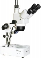
specialized, stereoscopic, optic, increase 10 – 160 x, плавное (зум-объектив), lens 1 – 4x, 2x, eyepiece WF10x, WF20x
children's, training, biological, optic, increase 64 – 1280 x, lens 4х, 10х, 40х(s), eyepiece WF16x, swivel eyepiece, Barlow lens, movable table
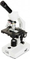
training, laboratory, biological, optic, increase 40 – 2000 x, lens 4x, 10x, 40x(s), 100x(s) oil, eyepiece WF10x/18 мм, WFH20x, movable table
training, laboratory, biological, optic, increase 40 – 2000 x, lens 4x, 10x, 40x, 100x, eyepiece 10х, 20х, swivel eyepiece, movable table
training, specialized, digital, increase 75 – 300 x, no eyepiece, camera, connection: AV-output, USB, card reader
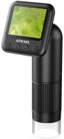
specialized, stereoscopic, digital, portable, increase 10 – 300 x, плавное (зум-объектив), camera, connection: AV-output, HDMI, USB, card reader
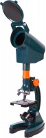
children's, biological, optic, increase 300 – 1200 x, lens 30х, 60х, 120х, eyepiece 10x, movable table
specialized, digital, portable, increase 400 – 800 x, плавное (зум-объектив), camera, connection: USB, card reader
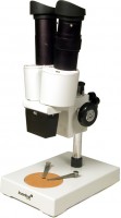
training, laboratory, stereoscopic, optic, increase 20 – 40 x, lens 2x, 4x, eyepiece WF10х
You might be interested in
Microscopes: specifications, types
Features
General purpose of a microscope.
Nowadays, there are 4 main options for the appointment: children's, educational, laboratory and specialized microscopes. At the same time, different options (at least from the first three) may well be combined in one model — for example, the simplest and most inexpensive educational microscopes may well be positioned as children's, and the most advanced as laboratory ones. Here is a detailed description of the different destinations:
— Children's. The most simple and inexpensive microscopes, designed primarily for children who are taking their first steps in the natural sciences (as well as for other undemanding users who do not need particularly advanced functionality). Accordingly, such devices lack advanced features such as focus lock, Keller lighting, video outputs (for digital and opto-digital models), a trinocular with the ability to connect a camera, etc. In addition, the body can be made in bright colours, and in plastic is usually used as the body material. However, many children's microscopes are equipped with turrets for quick re-tuning of magnification, and the total magnification factor may well exceed 600x out of the box and 1000x in the top configuration.
— Educational. Microscopes well suited for teaching applications; sometimes such an appointment is even di...rectly indicated by the manufacturer. The specific functionality of such models is quite diverse, the type can also be different (both biological and stereoscopic). In general, devices of this specialization occupy an intermediate position between simple and inexpensive children's microscopes and advanced laboratory equipment. At the same time, there are many models that have a combined purpose — "children's / educational" or "training / laboratory". The first variety is simple and inexpensive, for educational purposes it is suitable mainly for school; the second option, in turn, can be useful even at the university faculty of natural sciences.
— Laboratory. The most advanced type of modern microscopes, designed for full-fledged laboratory research and other serious tasks. Accordingly, such models are not cheap, but they provide a high-quality image and, in general, have the most extensive functionality (although the specific set of features, of course, may be different). Among the features found in laboratory microscopes are a movable stage, installation of light filters, 2 types of illumination (lower and upper), Keller illumination, suitability for special microscopy methods (fluorescence, phase contrast), etc.
— Specialized. Microscopes of a specific design and purpose, one way or another different from more traditional models. These differences may vary; accordingly, the specific specialization also differs. So, recently, portable models for smartphones have gained quite significant popularity: with the help of a special clothespin, such a device is attached directly to the gadget opposite the main camera, and the smartphone screen plays the role of an eyepiece. Another popular variety is compact digital microscopes without their own screens, connected to PCs or laptops via USB, and even to smartphones via Wi-Fi (including via the Internet). This also includes professional equipment with a fairly narrow specialization: stereoscopes with special mounts for dental prosthetics, for soldering microcircuits, etc.; microscopes for metallurgical research; devices on a tripod with a remote rod, designed to inspect individual areas on general objects; comparative microscopes for ballistic and trace investigations in forensics; and etc.
Type
- Biological. The term “biological” refers to microscopes designed for use primarily in biology and medicine - for studying cells, microorganisms and other similar objects of especially small size. One of the key differences between this type of microscopes and stereoscopic ones is the use of only one lens in the objective. As a result, the image turns out flat and it is impossible to estimate the volume of objects when looking through such a device. On the other hand, biological microscopes can provide a fairly high magnification factor - up to 2000x; and in tech areas where they are used, volumetricity is often not required.
Specialized microscopes, designated in the English-speaking environment by the term “Compound,” fall into the same category. Their strong point is the same large increase. Similar equipment is used in the metallographic field, pharmaceutical industry, jewelry, etc.
— Stereoscopic. Microscopes designed to obtain volumetric images (usually in overhead, reflected light). The traditional design of such a microscope includes a pair of lenses, paired eyepieces (binoculars), a relatively low magnification, a wide field of view, and a significant working distance (the distance from the lens to the object in question). Such features allow the user to observe a three-dimensional image with a good depth of field, and also, without any special obstacles, operate various tool...s in the field of view and clearly control their movements. This is indispensable for tasks such as repairing watches and other mechanisms, soldering microcircuits, creating miniatures, etc.
Note that this category may also include devices of a simpler design, with only one eyepiece and lens. Such a microscope is considered stereoscopic if it has a wide field of view, low magnification and a large depth of field; In terms of image quality, it will inevitably be inferior to models with two lenses, but will still provide a certain impression of three-dimensionality. Of course, such “pseudo-stereoscopes” are not suitable for serious tasks such as jewelry or watchmaking - most of them are intended for children and are designed so that a young researcher can examine in detail a pebble, flower or other object that is not shaped preparation for a biological microscope.
Operation principle
— Optical. Traditional microscopes, which work based on the use of lenses and other optical elements. They allow you to provide high image quality and a good magnification factor, while they do not depend on electricity (except that batteries may be needed for the backlight system). This type of microscope uses traditional eyepieces, but there are some models that allow you to connect an external camera and display the image on a computer display. Also note that this is the only principle used in stereoscopic models (see "Type")
— Digital. Microscopes of this type are actually digital cameras, supplemented with powerful magnifying optics. The image from such a camera must be displayed on the screen; some models have their own displays, others do not have displays and need to be connected to a computer/laptop. The advantage of the first variety is independence from external equipment, the advantages of the second option are compactness and relatively low cost. At the same time, it should be noted that in terms of magnification, most digital microscopes are inferior to optical ones, and this principle is not suitable for a stereoscopic image.
— Optical-digital. Microscopes that combine the features of optical and digital models (see relevant paragraphs). Such models differ from "purely digital" devices by more advanced optics, with a revolving head and a...high magnification; from optical ones — with a built-in camera and using a screen as an eyepiece (traditional eyepieces are not used in optical-digital models).
Magnification
The range of magnifications provided by the device is from minimum to maximum.
The magnification of the microscope is calculated by the formula "the magnification of the eyepiece is multiplied by the magnification of the objective." For example, a 20x objective with a 10x eyepiece will give a magnification of 10*20 = 200x. Modern microscopes can be equipped with multi-objective turrets, zoom lenses (see below) and interchangeable eyepieces — so that in most models the magnification can be adjusted. This allows you to adjust the device to different situations: when you need to consider small details, a high degree of magnification is used, but to expand the field of view, the magnification must be reduced.
Detailed recommendations on optimal multiplicities for different tasks can be found in special sources. Here we note that many manufacturers go to the trick and indicate the maximum value of the magnification by the degree of magnification achieved with an additional Barlow lens. Such a lens can indeed give a serious increase in magnification, but it is not a fact that the image will turn out to be of high quality; for more details, see "Complete set".
Research method
Research methods applicable to this microscope model.
— Bright field. The most famous and widely used method of light microscopy. The object under consideration in such studies is placed on a light background, against which it looks darker. Note that different methods of illumination can be used for research: direct through, oblique, reflected. The first option (when the light from a lamp or a mirror under the stage shines through the sample) is optimal for studying transparent samples, the key details of which are darker than the general background. Typical examples are thin sections of animal and plant tissues. Oblique light is similar in application specifics, while it gives a grey background and is inferior to direct light in terms of backlight efficiency, but provides a more embossed image. As for reflected light, in this case it is indispensable when examining opaque objects: samples of ores and other materials, semiconductor wafers, etc. Anyway, bright-field microscopy well reveals, first of all, details that are noticeably different in light transmission or refractive index from the surrounding background (with through illumination), or give noticeable reflections / shadows (with reflected light).
— Dark field. A kind of opposite of bright-field research: the object under consideration or its individual elements are lighter than the surrounding background. However, this is not just a “negative” of the image, but a separate method with its own c...haracteristics. Illumination in dark-field microscopy is usually through, but it is carried out in a specific way: the middle of the light beam is blocked by a hood, and the light “cylinder”, passing through the condenser lens, turns into an “hourglass”. At the same time, in the narrowest place of such a "clock" there is a preparation, and towards the lens, the light cone expands so that it does not fall into the optics. Thus, the user sees in the microscope only the light scattered by the preparation and the dark background around. This method of research, among other things, allows you to identify "smooth" details that do not stand out sharply against the surrounding background and are not visible in a bright-field study. Among the applications of dark-field microscopy are the work with unstained biological preparations (cells, tissue samples, microorganisms), as well as the study of some transparent materials for small surface defects.
— Phase contrast. A method used to study transparent and colourless objects with an inhomogeneous structure, used when this inhomogeneity cannot be detected by more traditional bright-field microscopy. The idea of this method is that when passing through structures with different refractive indices, light receives different phase changes. These changes are not visible with ordinary optics, but they can be made visible with the help of special equipment — namely, a condenser and a lens of a special design. Accordingly, such equipment is necessarily included in the scope of delivery of the microscope.
— Fluorescent. This method provides the illumination of observed objects with light of a certain wavelength, under the influence of which these objects or their individual elements begin to glow, while the background remains dark. If necessary, coloring substances are introduced into the preparation to improve luminosity (a typical example is biological objects, most of which fluoresce rather weakly by themselves). For illumination, usually, UV radiation is used, therefore this method is also called ultraviolet microscopy; The image enters the eyepiece of the microscope through a filter that filters out UV rays, but freely passes the visible glow of the drug.
One of the main features of fluorescence microscopy is high resolution: it allows you to clearly see even very small objects that are not visible in the usual visible range. In fact, this method in terms of resolution is between classical optical and electron microscopy; At the same time, in contrast to electron and atomic microscopes, devices with the support of the UV method make it possible to examine even the hardware of living cells and microorganisms. And some special variants of this technique make it possible to achieve not micro, but nanoscopic magnifications. The second popular use of fluorescence microscopy is the detection of particles, elements, inclusions, etc., which are not visible under ordinary light, but stand out well in ultraviolet light. A typical example is the surface of many metals and alloys.
Portable
This category includes small microscopes, originally designed for the possibility of constant carrying with you and use "in the field", outside of laboratories. Some of these devices are comparable in size and weight to flashlights. The multiplicity of portable microscopes is small — up to 100 – 200x, in some models up to 500x; however, a high degree of magnification is not required in this application. Such devices are valued by jewelers, forensic experts and other professionals who often have to conduct research in the field.
Inverted
In inverted microscopes, the mechanism is arranged “upside down”: the lens is located under the object stage, and the illumination system is installed on top (however, its presence is not necessary). The location of the eyepiece does not fundamentally differ from the traditional design, the image is transmitted to it through a system of prisms. In addition, long-focus objectives are used in such models, allowing a cover slip thickness of 1.5 mm or even more (in conventional microscopes, the allowable thickness is usually 0.17 mm).
This design provides a number of advantages over the traditional one. First, from below, in most cases, it is most convenient to view the contents of Petri dishes and other dishes with a transparent bottom. Secondly, the reference and working (considered) surfaces of the observed object coincide with such a design. Here it is worth recalling that the working surface of the preparation for optimal visibility should be perpendicular to the optical axis of the lens, while the supporting surface is always perpendicular to it. So in inverted microscopes, there is no need to waste time on additional adjustment of the preparation position. Thirdly, such devices are suitable for working with objects that are very significant in height; and in those models where there is no lighting system or it is removed, there are no height restrictions at all (the main thing is that the object table can withstand the weight o...f the object). The main disadvantages of inverted microscopes are complexity and, accordingly, higher cost compared to classical counterparts.
This design provides a number of advantages over the traditional one. First, from below, in most cases, it is most convenient to view the contents of Petri dishes and other dishes with a transparent bottom. Secondly, the reference and working (considered) surfaces of the observed object coincide with such a design. Here it is worth recalling that the working surface of the preparation for optimal visibility should be perpendicular to the optical axis of the lens, while the supporting surface is always perpendicular to it. So in inverted microscopes, there is no need to waste time on additional adjustment of the preparation position. Thirdly, such devices are suitable for working with objects that are very significant in height; and in those models where there is no lighting system or it is removed, there are no height restrictions at all (the main thing is that the object table can withstand the weight o...f the object). The main disadvantages of inverted microscopes are complexity and, accordingly, higher cost compared to classical counterparts.
Turret
The number of lenses in the microscope turret.
The turret is a round nozzle with several lenses of different magnification. By turning such a nozzle, you can change the lens currently used; and the more lenses, the wider the choice for the user when choosing the optimal magnification of the microscope. On the other hand, numerous optics affects the dimensions and price of the device. In light of this, most modern microscopes have 3-4 lenses — this number is considered optimal in terms of functionality and price.
The turret is a round nozzle with several lenses of different magnification. By turning such a nozzle, you can change the lens currently used; and the more lenses, the wider the choice for the user when choosing the optimal magnification of the microscope. On the other hand, numerous optics affects the dimensions and price of the device. In light of this, most modern microscopes have 3-4 lenses — this number is considered optimal in terms of functionality and price.
Lens
— Zoom lens. Lens with variable magnification. Such optics allow you to smoothly change the overall magnification of the microscope within certain limits, without changing the objective/eyepiece and without even looking up from observations. On the other hand, zoom lenses are more complicated and more expensive than constant magnification optics. Therefore, they are mainly used in stereoscopic microscopes (see "Type"): in the repair, assembly and other tasks for which such devices are used, the ability to smoothly adjust the multiplicity is extremely useful.
— magnification factor. The magnification provided by the lens. This parameter, along with the magnification of the eyepiece, affects the overall magnification of the device (see above). Most biological microscopes (see "Type") are equipped with several different magnification objectives on the turret; this allows you to adjust the degree of magnification as desired by the user. The standard magnification options for such lenses are 4x, 10x, 40x, 100x.
— Achromat. One of the varieties of colour correction used in lenses. The need for colour correction is due to the fact that light of different colours is refracted differently by lenses, and without additional measures, the image in the microscope would be blurred with iridescent stains. Achromatic is one of the simplest types of colour correction; in such optics, colour distortions in yellow and green are corrected.... Achromatic lenses have simple design and low cost. However the image quality in them is far from perfect: such a lens gives a clear image only in the centre of the image, the width of the sharpness zone is about a third of the total width of the field of view, and red-blue streaks may appear along the edges of the image. However, this is quite enough for general acquaintance, initial training, and often for more serious tasks.
— Planachromat. An improved and improved version of achromatic lenses (see above). Plan achromats provide additional correction of the field curvature, due to which the area of a clearly visible image in such lenses is at least 2/3 of the total width of the field of view, and often even more. It is these lenses that are recommended for serious study and professional use.
— Rim diameter. The size of the thread used to mount the lens. A larger bore usually means a wider lens, which means higher aperture and better image quality. On the other hand, the large size affects the dimensions, weight and cost of optics. In modern microscopes, diameters from 20 to 35 mm are mainly found. Knowing the size of the thread, you can purchase replacement or spare lenses for the device.
Eyepiece
— Monocular. An eyepiece with a single lens that can only be viewed with one eye. For obvious reasons, it is only used in biological microscopes (see "Type"). The advantages of monoculars are primarily smaller size and cost than other varieties; in addition, they do not require adjustment for interpupillary distance. On the other hand, constantly looking into the eyepiece with one eye is tiring, so this option is poorly suited for situations where you have to look into the microscope often and for a long time.
— Binocular. Dual eyepiece that can be viewed with both eyes at once. Note that such optics are used not only in stereomicroscopes, originally intended for viewing an object through two lenses (see "Type"), but also in biological microscopes with one lens. The fact is that looking into an optical device with two eyes is much more convenient than with one, while the eyes are less loaded and fatigue does not occur so quickly. Therefore, for serious tasks associated with frequent use of a microscope, binoculars (or trinoculars, see below) are the best option. Such optics cost more than monocular, but this is offset by ease of use.
— Trinocular. A kind of binocular (see the relevant paragraph), supplemented by a third optical channel for a special camera-video eyepiece. Such a camera is usually connected to a PC or laptop; by installing it in the soc...ket for the third eyepiece, you can take photos and videos, as well as display the image in real time on the computer screen. At the same time, you can look through the microscope in the usual way. Devices with trinoculars are very functional and versatile, but they are complex and expensive.
— LCD screen. The microscope has an LCD screen that replaces the traditional eyepiece. You do not need to bend over to such a device each time to view the image, which is very convenient if observations need to be combined with record keeping and other similar activities. Microscopes of this design usually have a photo and video function, as well as various built-in tools — for example, a scale grid for estimating the size of visible objects, displayed directly on the screen. In addition, the image on the screen can be seen not only by the direct user, but also by everyone who is nearby; such features are indispensable during training sessions, consultations, presentations, etc. On the other hand, such microscopes turn out to be bulky and expensive.
— magnification factor. The magnification provided by the eyepiece. This parameter, along with the lens magnification, affects the overall magnification of the device (see above). The classic option for eyepieces in microscopes is 10x, but higher values \u200b\u200bare also found. The package may include several eyepieces, of different magnification — to change the overall degree of magnification. There is a multiplicity designation with a letter index, for example, WF10x. This means that the eyepiece has an extended field of view (WF — wide, EWF — extra wide, UWF — extra wide).
— Eyepiece tilt. The tilt of the eyepiece determines the position of the observer's head when looking through the microscope and the overall usability. According to this indicator, three main options can be distinguished: fixed angle, adjustable angle, without tilt. The fixed angle is most often 30° or 45° relative to the horizontal, these values are considered the most convenient. In angle-adjustable microscopes, the entire stand, with tube and stage, is fixed to the base with a swivel mount. This is the most convenient option, allowing you to adjust the tilt to your preference, but the mount tends to become loose over time, so it is rarely used in professional microscopes. The third variety — vertical microscopes, without tilt — have not received much distribution: this design is used in some stereoscopic models (see "Type") in order to ensure that the stage remains strictly horizontal (this is important for some work with microscopic objects).
— Rim diameter. The nominal diameter of the eyepiece used in the microscope, as well as the diameter of the hole in the tube, designed to install the eyepiece. Several standard diameters are used in modern microscopes, in particular 23 and 27 mm. In fact, this parameter is necessary, first of all, if you plan to purchase spare or replacement eyepieces for the microscope, or if you already have an eyepiece on the farm, and you need to evaluate its compatibility with this model.
— Diopter adjustment. The range of diopter correction provided in the eyepiece. This correction is used so that a nearsighted or farsighted person can look through the microscope without glasses or contact lenses. In most models with this function, the correction range is about 5 diopters in both directions; this allows the microscope to be used for low to moderate myopia/farsightedness.
Rotary eyepiece
This feature means that the eyepiece that the microscope is equipped with can rotate around a vertical axis — in other words, right and left. Typically, the range of rotation is a full 360°.
The swivel head of the eyepiece does not affect the main features and capabilities, but provides additional convenience for the user: the eyepiece can be rotated to the optimal position depending on the situation. This can be useful, for example, when two students or laboratory assistants sitting next to each other use one microscope with a preparation for two — if necessary, each can turn the eyepiece towards him without moving the entire device. The reverse side of this advantage is some complication of the design and an increase in its price.
The swivel head of the eyepiece does not affect the main features and capabilities, but provides additional convenience for the user: the eyepiece can be rotated to the optimal position depending on the situation. This can be useful, for example, when two students or laboratory assistants sitting next to each other use one microscope with a preparation for two — if necessary, each can turn the eyepiece towards him without moving the entire device. The reverse side of this advantage is some complication of the design and an increase in its price.
Interpupillary distance
Interpupillary distance in a microscope equipped with an eyepiece "under two eyes" — a binocular or trinocular.
In fact, this paragraph indicates the distance between the optical centers of the eyepieces. For normal visibility, it must exactly match the distance between the pupils of the user's eyes — hence, among other things, the name "interpupillary". And since the distance between the pupils can vary markedly for different people, then in all modern microscopes (for which this is generally relevant), the eyepieces are made movable, and the width of their location can be adjusted. In this paragraph, respectively, the range of such adjustment is indicated. Most often, it ranges from 55 to 75 mm — this is quite enough to choose the option for almost any adult user. But there are also more extensive adjustment ranges, mainly with an extension to the smaller side — for example, 52 – 76 mm or 48 – 75 mm. Such characteristics may be useful, in particular, when it comes to a children's microscope.
In fact, this paragraph indicates the distance between the optical centers of the eyepieces. For normal visibility, it must exactly match the distance between the pupils of the user's eyes — hence, among other things, the name "interpupillary". And since the distance between the pupils can vary markedly for different people, then in all modern microscopes (for which this is generally relevant), the eyepieces are made movable, and the width of their location can be adjusted. In this paragraph, respectively, the range of such adjustment is indicated. Most often, it ranges from 55 to 75 mm — this is quite enough to choose the option for almost any adult user. But there are also more extensive adjustment ranges, mainly with an extension to the smaller side — for example, 52 – 76 mm or 48 – 75 mm. Such characteristics may be useful, in particular, when it comes to a children's microscope.
Maximum working distance
The greatest working distance provided by the microscope.
The working distance is the distance from the lens to the object in question. This parameter is important primarily for stereomicroscopes (see "Type"): the more space remains under the lens, the more convenient it is to work with various tools and devices in the field of view of the device. However, here it should be taken into account that the maximum working distance is achieved at the minimum magnification factor, as the magnification increases, the lens has to be brought closer to the object in question. For biological microscopes, the working distance does not really matter: such devices work mainly with flat preparations, to which the lens can be brought almost close.
The working distance is the distance from the lens to the object in question. This parameter is important primarily for stereomicroscopes (see "Type"): the more space remains under the lens, the more convenient it is to work with various tools and devices in the field of view of the device. However, here it should be taken into account that the maximum working distance is achieved at the minimum magnification factor, as the magnification increases, the lens has to be brought closer to the object in question. For biological microscopes, the working distance does not really matter: such devices work mainly with flat preparations, to which the lens can be brought almost close.
Object table
The design of the object stage provided in the microscope.
— Stationary. Subject table, fixed motionless; focus in such microscopes is carried out by moving up and down the tube with the objective and the eyepiece. Such systems are simple and inexpensive, but focus while looking through a constantly moving eyepiece is not very convenient. In addition, for advanced biological microscopes (see "Type") with binoculars and trinoculars (see "Eyepiece"), this option is also poorly suited for some design reasons. But the vast majority of stereomicroscopes are equipped with stationary tables — this is the most reasonable design, taking into account the specifics of the application.
— Movable. In microscopes of this type, the entire optical system is fixedly fixed on a tripod, and the stage can be moved up and down to focus the optics. This design is found exclusively in biological microscopes (see "Type"). It is somewhat more complicated and expensive than with a fixed table, but at the same time it is much more convenient: when focus, the eyepiece does not move, which allows you to comfortably adjust the image without looking up. In addition, it is the movable stage that is most suitable for advanced devices with binoculars and trinoculars (see "Eyepiece"), almost all such microscopes have such equipment.
Drug agent
Presence of a preparation agent in the design of the object table.
The preparation guide is a device for smooth movement of slides under the microscope lens, as well as fixing the conditional coordinates of individual sections of the preparation. Mechanisms are responsible for the movement, allowing the glass to be shifted separately in the longitudinal and transverse directions. Coordinate fixation is provided by special scales with verniers, the accuracy of determining coordinates can be from 0.1 to 0.01 mm.
This feature is found exclusively in biological microscopes (see "Type"). Its presence can be extremely important for studies involving high magnification factors. Without a slider, the glass would have to be moved by hand, and finding certain areas would be a very difficult, if not impossible, task.
Focus
Types of focus (focus) provided in the microscope. Focus is carried out by changing the distance between the object under consideration and the lens; its types can be:
— Rough. This method means that there is one rotary control responsible for moving the lens or stage. The advantages of this design are simplicity and low cost. At the same time, focus at high magnifications in such microscopes is a rather difficult task: you have to turn the tuning knob literally in fractions of a millimetre.
— Coarse / Fine. Focus, carried out by two mechanical controls — for preliminary focus and for final fine tuning. Such a tuning is more convenient in itself than only a rough one (see above), and at high magnifications it can be simply irreplaceable. On the other hand, the presence of an additional regulator complicates and increases the cost of the design, so this option is found mainly in semi-professional and professional microscopes.
— Manual. A method that assumes the absence of a focus mechanism as such. Focus in such devices is carried out due to the fact that the user manually moves the lens — for example, moving it up and down on a vertical tripod and fixing it in the desired position with a clamp, or tilting it back and forth on a swivel mount. This option is only suitable for models with a low magnification that do not require special accuracy when focus; it is found mainly in digital microscopes without thei...r own screen (see "How it works"), as well as portable models (see the relevant paragraph).
Focus lock
Possibility to block the focus mechanism of the microscope. One way to use this feature is to work with numerous the same type of specimens: by blocking the focused microscope, you can change specimens without wasting time focus at each change. In addition, blocking will not interfere when working at very high magnifications (from 1000x and higher). Focus on such magnifications must be very precise, and the working distance is small — as a result, accidentally hitting the coarse focus knob, you can thoroughly knock down the settings or even “drive” the lens into the preparation. Blocking helps to avoid such troubles.
Backlight
The type of stage illumination used in a microscope.
- Light-emitting diode (LED). The most advanced type of illumination to date. The LEDs produce a bright white light with a cool colour, perfect for working with transparent samples. Such light sources can be equipped with dimmers. In addition, LED backlighting is extremely economical in terms of energy consumption and generates almost no unnecessary heat. All this makes this option suitable even for the most advanced microscopes.
— Halogen. Before the advent of LEDs, this type of illumination was the main choice used in biological microscopes (see "Type") of the intermediate and professional levels. Halogen lamps provide a powerful stream of light, while the brightness of the backlight, usually, can be adjusted; the emission spectrum turns out to be quite convenient for observations, and the heating is relatively small (although more than in LEDs). In terms of energy efficiency, such lighting is inferior to LED, but surpasses incandescent lamps.
- Incandescent lamp. The most simple and inexpensive type of backlight. Actually, it is the low cost that is the main advantage of such systems. But the disadvantages of incandescent lamps are many. Firstly, they give a warm shade of glow that distorts colour reproduction; for simple tasks this is not critical, but in serious studies it is unacceptable. Secondly, the lamp gets very hot, which can adversely affect the drug. Thirdly, such lighting c...onsumes quite a lot of energy. As a consequence, incandescent lamps are found exclusively in inexpensive, entry-level microscopes, and even among these, they are slowly falling into disuse.
- Mirror. Lighting with a mirror that reflects light from a window, ceiling lamp, or other external light source. The advantages of this option include simplicity, low cost, compactness and complete independence from energy sources. On the other hand, such a microscope depends on external illumination, and setting up a mirror requires certain skills and can be quite difficult to get used to. Therefore, in its pure form, mirror systems are used relatively rarely, however, a mirror can be provided as an addition to another source of illumination, for example, a halogen lamp.
Top illumination
The presence in the microscope of illumination directed from top to bottom. In biological microscopes (see "Type"), such illumination makes it possible to examine opaque objects; also in some cases it can be used as an addition to the main lower lighting. And in stereoscopic models, only overhead light is used — this is due to the specifics of the application.
Bottom illumination
Bottom lighting is a lighting system, the light from which is directed from the bottom up.
In conventional (not inverted) microscopes, such illumination is directed towards the objective through a hole in the stage. It is this type of illumination that is used for classical bright-field microscopy using through illumination; Thus, the lower arrangement of the illumination is traditional for biological microscopes and is provided for in most of these models. But the presence of this function in "stereoscopes" is not typical, although it also occurs.
In turn, in inverted microscopes, the upper and lower illuminations are actually “swapped”. Accordingly, in such models, this function is intended for examining preparations (mostly opaque) in reflected light, and the light flux is directed from the lens to the preparation.
Condenser
Features of the design of the condenser installed in the microscope.
The condenser is part of the illumination system in biological microscopes (see "Type"). This is an optical system that processes the light flux entering the preparation glass in a special way. Different situations may require different ways of doing this; accordingly, different types of condensers can be used in microscopes. However, the most popular nowadays is the simplest Abbe condenser. It ensures the concentration of the beam of light and its uniform distribution over the field of view. Initially, such a device was intended for studies using the bright field method, but it can also be used for phase-contrast observations. The Abbe condenser can be equipped with an iris aperture diaphragm — with its help you can reduce the brightness of the illumination — as well as colour filters.
Other, more specific types of condensers (for example, phase or dark field) are usually purchased separately and are rarely included in the standard microscope equipment.
The characteristics of the condenser may indicate NA — the size of the aperture (active hole) in millimetres, for example, NA \u003d 1.2. This is a rather specific setting; suffice it to say that it is selected by the manufacturer for complete lenses and does not fundamentally affect the choice of a microscope.
Diaphragm
The type of diaphragm installed in the microscope.
The diaphragm is a device that partially blocks the flow of light from the microscope lighting system. It is used mainly for adjusting lighting, as well as for some more specific tasks (in particular, changing the depth of field). When adjusting the diaphragm, the size of its working opening changes - and, accordingly, the actual light transmission; and different types of diaphragms ( iris or rim) differ in adjustment features:
- Iris. The name comes from the Latin word for the iris of the eye - similar devices work on a similar principle. The iris diaphragm consists of a set of specially shaped blades (the so-called lamellas). When moving to close, these petals move from the edges of the working hole to the center, reducing its size; when opening, they correspondingly move outward. Iris diaphragms are more complex and more expensive than rim diaphragms, but they have a number of important advantages over them. First of all, the light transmission throughout the entire operating range of such devices changes smoothly, which allows you to select the settings as accurately as possible. You can manage the settings without interrupting your monitoring of the drug; At the same time, iris diaphragms are also as compact and lightweight as possible. As a result, this option is the most popular in microscopes of the middle class and above, and...is also often found even in simpler models.
- Disk. Another name is revolver. This type of diaphragm is a rim with holes of different sizes made in it; By rotating the rim, you can place different holes in the field of view of the microscope and, thus, change the light transmission. The main advantages of such devices are simplicity of design, low cost, reliability and ease of repair. On the other hand, disc diaphragms are less practical and sophisticated than iris diaphragms - in particular, they are very bulky and do not allow for smooth adjustment. In light of this, this option is used mainly among entry-level microscopes, where advanced characteristics are not required - and an affordable price, on the contrary, is of key importance.
Light filters
The presence of light filters in the scope of delivery of the microscope.
Light filters are installed in the lighting system; they can be interchangeable or built-in (usually on a turret). Anyway, such devices change the characteristics of light, adjusting it to the specifics of the situation. The types and purpose of light filters can be different, as well as their range in the kit; here are some of the most common options:
— Blue colour. Useful in cases where light from an incandescent or "halogen" lamp is used for illumination. Such a filter equalizes the colour temperature (white balance), making the shades of colours colder and providing natural colour reproduction; this is especially important for microphotography, since a properly set white balance is critical to obtaining high-quality images.
— Yellow colour. Kind of the opposite of blue: lowers the colour temperature, giving the image a warmer tint. It is also sometimes useful for adjusting white balance, but yellow filters have another important use: they are well suited for detecting imperfections in metallic surfaces.
— Green colour. Achromatic and planachromatic objectives, which are installed in most modern microscopes, are best at eliminating aberrations in the green part of the spectrum. With this in mind, similar filters are applied: an image painted in a green tint has the least visible distortion. In addition, most objectives...for phase contrast microscopy are also most effective in the green part of the spectrum (although exceptions are possible).
— Matte (diffuser). White colour filters that do not change the colour of the light, but provide additional dispersion. This can be useful, in particular, when working with low magnification lenses.
— Neutral. Filters in different shades of grey. Used to reduce the intensity of lighting without changing its other characteristics. Such devices can be especially useful when taking photographs — namely, if the camera does not have a sufficiently fast shutter speed. Note that a similar effect can be achieved using a microscope diaphragm, but this is not always the best option when shooting. So, narrowing the aperture reduces the field of view and increases the depth of field (the latter is also not always desirable), while filters do not affect these parameters; besides, in some situations, even the narrowest aperture may not be “dark” enough.
— Light filters for coloured preparations. Improve the visibility of elements painted in a particular colour. Such fixtures are especially popular in biological studies, as they are the most commonly stained specimens and are also the most susceptible to dye fading, making it difficult to view under normal lighting conditions. Note that filters of this type, in contrast to the colour filters described above, do not colour the entire image in a certain colour, but only muffle all other colours, except for their “native”.
— Fluorescent. Filters used in fluorescence microscopy. They are divided into two types — exciting (they separate UV radiation from the general backlight spectrum to illuminate the drug) and closing (protect the user's eyes from ultraviolet radiation and at the same time let the fluorescent glow of the drug pass through).
Integrated camera
The presence in the microscope of its own built-in camera, which allows for photo and video shooting of objects in the field of view, as well as displaying the image on an external screen (or your own, if available). Specific features of the application of this function may be different, depending on the design features. So, some microscopes (mostly portable, see the relevant paragraph) work only with external screens, others have their own displays, others can work with their own and with an external screen. Similarly, the features of photo / video recording may vary; see the relevant paragraph for more details.
Number of megapixels
Camera sensor resolution in megapixels (millions of pixels).
The higher the resolution of the matrix, the higher the video resolution can be (see below), the more detailed the image is capable of providing the camera. At the same time, note that as the number of megapixels increases (without changing the size of the matrix), the size of each individual pixel decreases, which increases the likelihood of noise and deterioration of the overall image quality. Therefore, in itself, high resolution is not necessarily a sign of high quality — a lot depends on other points, for example, on the size of the matrix.
The higher the resolution of the matrix, the higher the video resolution can be (see below), the more detailed the image is capable of providing the camera. At the same time, note that as the number of megapixels increases (without changing the size of the matrix), the size of each individual pixel decreases, which increases the likelihood of noise and deterioration of the overall image quality. Therefore, in itself, high resolution is not necessarily a sign of high quality — a lot depends on other points, for example, on the size of the matrix.
Video resolution
The maximum video resolution that the microscope camera can capture.
The higher the resolution of the video, the more details you can see on it, the less details will be blurry. On the other hand, high resolution means large volumes of video materials, which, accordingly, requires capacious media for their storage and fast communication channels for broadcasting video in real time. Yes, and this indicator affects the cost.
The higher the resolution of the video, the more details you can see on it, the less details will be blurry. On the other hand, high resolution means large volumes of video materials, which, accordingly, requires capacious media for their storage and fast communication channels for broadcasting video in real time. Yes, and this indicator affects the cost.
Features
— Adjustment of interpupillary distance. The ability to change the distance between the eyepieces in a binocular or trinocular microscope (see "Eyepiece"). For normal visibility, it is necessary that the distance between the lenses of the eyepieces correspond to the distance between the pupils of the user. This distance varies from person to person, so this setting may be required for comfortable use.
— Brightness adjustment. The ability to change the brightness of the backlight — to adjust the lighting to the specifics of the situation. For example, to study a thin transparent preparation in a bright field, high brightness will be unnecessary, but when transilluminating a dense dark object, it is indispensable.
— Illumination according to Keller. The presence of illumination in the microscope according to the Keller system. Such lighting is used exclusively in biological models (see "Type"), it is a sign of a professional level device. The Keller system complicates and increases the cost of the design, in addition, it may require specific settings, but with the right settings, the quality of the lighting is very high, and the image is as reliable as possible. Note that in microscopes there is a so-called. "simplified Keller system", when the settings are set at the factory and cannot be changed; however, in this case, it is precisely the full-fledged,...adjustable Keller lighting that is meant.
— Photo / video recording. Possibility of photo and video filming of the image visible in a microscope. Features of the implementation of this function in different microscopes may be different. For example, some models need to be connected to a computer, while others can record materials directly to a memory card or other media. Also, the cameras themselves, carrying out the shooting, can be both built-in and removable (see "Packaging" / relevant paragraphs).
Connection interfaces
Ways to transfer data to other devices provided in the design of the microscope.
This parameter is relevant primarily for digital and opto-digital models, as well as for individual optical devices equipped with cameras. All described microscopes can be equipped with AV and HDMI outputs, universal USB ports, removable media card readers, and Wi-Fi wireless modules. Here is a detailed description of each interface:
— AV output. Analogue output for video signal transmission. It is used primarily for live transmission of images from a microscope camera, and in some models — also for viewing footage stored in memory. Such outputs do not support HD resolutions and, in general, are inferior to HDMI in terms of overall “picture” quality (with the same camera characteristics). On the other hand, specifically for microscopes, these moments are not so often critical; analogue connectors are still quite popular in both conventional video equipment and special equipment; and the implementation of this interface is inexpensive. Therefore, AV outputs can be found even in fairly advanced models.
— HDMI. Digital output for video signal transmission. Similarly, AV can be used both for real-time broadcasting and for using the microscope as a video player when viewing saved materials (if such...a possibility is provided for in this model at all). At the same time, such outputs are more advanced than analogue AV: HD-quality images (including Full HD and higher) can be transmitted via HDMI, and the signal is very resistant to interference. We also recall that this interface is extremely common in modern video technology — in particular, the presence of at least one HDMI input is almost mandatory for TVs and monitors that support HD standards. On the other hand, the implementation of HDMI is noticeably more expensive, and it makes sense to use it with fairly advanced cameras, which in themselves significantly affect the price of microscopes. Therefore, such outputs can be found mainly in rather expensive and advanced devices.
— USB. Universal connector that allows different applications; a specific set of these options is directly related to the functionality of the microscope. Typical examples of using USB include the following: copying captured photos and videos to a computer or laptop; live image broadcast; remote control via PC / laptop (for example, moving the parent drug); charging the built-in battery, etc. The specific type of USB connector in the microscope may vary, however, usually, an appropriate cable is supplied in the kit for connecting to a standard full-size port.
— Card reader. The device for working with memory cards is usually SD, and in miniature pocket models — microSD. Such cards usually contain materials captured by the camera. In general, this function makes it much easier to copy information to other devices that also have card readers — primarily laptops and PCs; and miniature microSD cards are also supported by smartphones, tablets and other portable gadgets. Anyway, removing the card from the microscope and installing it in another device is often easier and faster than fiddling with a wired or Wi-Fi connection.
— Wi-Fi. A wireless module, which in this case is mainly used to communicate with an external device — such as a smartphone, laptop or PC. A Wi-Fi connection allows you to at least broadcast the image from the camera and copy the photos taken by it, and often also control other functions and settings (light brightness, movement of the driver, etc.). At the same time, the absence of wires provides additional freedom of movement and overall convenience. However, note that the specific communication format may be different, it should be specified separately. So, some models support only direct connection over a relatively short distance (in fact, up to a couple of tens of metres, or even less). Others are able to connect to an external device via the Internet, and here the distance does not play a role — there would be access to the World Wide Web. Still others allow both formats of work. Also note that individual devices with this function do not have their own screens at all and are designed for use with external gadgets; This design makes the microscope as compact and easy to carry as possible.
Power source
Methods of nutrition provided in the microscope. Even optical models may require a power source to run the backlight (see above), while for other varieties, power is almost a must. Some models may support multiple power types.
— 230 V network. Connection to a regular 230 V socket. Quite a convenient and practical option, only poorly suitable for portable models (see above).
— USB port. Power supply from the USB connector is often found in digital microscopes (see "How it works"): the device is powered from the same connector through which it is connected to a computer or other external screen. And in optical models, such power supply can be provided in addition to the 230 V network described above. Note that USB ports, among other things, are also found in laptops and other portable devices, which makes it possible to use such microscopes even if there are no outlets nearby. This is especially useful for portable devices (see above).
— Accumulator. Powered by its own built-in battery, in some cases — non-removable. This option makes the microscope completely autonomous and allows you to use it even in the complete absence of external power sources nearby. On the other hand, this moment is relevant mainly for portable models, and then only in some cases, and the built-in battery noticeably affects the weight, dimensions and price of the device. Therefore, purely cordless microscopes are extremely rare, more often this method of power supply is pro...vided in addition to the 230 V network or USB (see above) — as a spare in case of problems with external power.
— Batteries. Another type of autonomous power supply, along with the batteries described above. The presence of a battery compartment is cheaper than the built-in battery, but the batteries themselves have to be purchased separately — and either regularly buy disposable cells, or pay a rather large amount for batteries and a charger for them. In addition, the quality of batteries is highly dependent on the specific brand, and not all cells can normally “start” the microscope and provide an acceptable battery life. Therefore, such power, like battery power, is rare in its pure form, more often it complements the connection to a 230 V network or USB.
In box
Additional equipment supplied with the microscope.
— Camera. In this case, we mean a removable camera installed either on the main optical channel (to use an external screen as an eyepiece), or on the third additional channel of the trinocular (see "Eyepiece"). In addition, there are also built-in cameras (see the relevant paragraph). Some models supplied without a camera allow you to purchase it separately, but this configuration option is generally more convenient.
— Adapter for smartphone. A device that allows you to install a smartphone on a microscope so that the camera of the device “sees” the image in the eyepiece. Thus, you can take photos and videos on your smartphone, as well as use its screen as an eyepiece — for example, if you want to show the image to several people at once.
— A set of accessories and preparations. A set of accessories for working with a microscope. Such a set usually includes at least slides and coverslips; in addition to them, the kit can be supplied with preparation tools, various auxiliary compounds (resin for gluing, oils and liquids for immersion lenses), as well as ready-made preparations for testing the capabilities of the microscope and initial training in working with it.
— Lens Barlow. An additional lens that is installed in front of the eyepiece and changes the overall magnification — usually upwards, but vice versa is...also possible. To calculate the overall magnification when using such optics, you need to multiply the initial magnification of the device by the magnification of the lens: for example, a 200x microscope with a 1.6x Barlow lens will give 200 * 1.6 = 320x magnification. This is partly why Barlow lenses have a very low magnification — even it gives a significant increase in magnification. The second reason is that it makes sense to increase the overall magnification only up to a certain limit — above this limit, the optics will only stretch the image without increasing the detail. Actually, in many microscopes, this is exactly what happens if you set the device to the maximum magnification and install a Barlow lens. So this device should be considered more as a tool for adjusting the magnification at medium magnification, and not as a way to increase the maximum magnification.
— Cover / case. Case for storage and transportation of the microscope. Covers are called soft cases, they are mainly designed to protect against pollution; cases are made of hard materials, they are more bulky, but they are also able to protect the device from shocks and shocks.