Features
General purpose of a microscope.
Nowadays, there are 4 main options for the appointment:
children's,
educational,
laboratory and
specialized microscopes. At the same time, different options (at least from the first three) may well be combined in one model — for example, the simplest and most inexpensive educational microscopes may well be positioned as children's, and the most advanced as laboratory ones. Here is a detailed description of the different destinations:
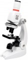
— Children's. The most simple and inexpensive microscopes, designed primarily for children who are taking their first steps in the natural sciences (as well as for other undemanding users who do not need particularly advanced functionality). Accordingly, such devices lack advanced features such as focus lock, Keller lighting, video outputs (for digital and opto-digital models), a trinocular with the ability to connect a camera, etc. In addition, the body can be made in bright colours, and in plastic is usually used as the body material. However, many children's microscopes are equipped with turrets for quick re-tuning of magnification, and the total magnification factor may well exceed 600x out of the box and 1000x in the top configuration.
— Educational. Microscopes well suited for teaching applications; sometimes such an appointment is even di
...rectly indicated by the manufacturer. The specific functionality of such models is quite diverse, the type can also be different (both biological and stereoscopic). In general, devices of this specialization occupy an intermediate position between simple and inexpensive children's microscopes and advanced laboratory equipment. At the same time, there are many models that have a combined purpose — "children's / educational" or "training / laboratory". The first variety is simple and inexpensive, for educational purposes it is suitable mainly for school; the second option, in turn, can be useful even at the university faculty of natural sciences.
— Laboratory. The most advanced type of modern microscopes, designed for full-fledged laboratory research and other serious tasks. Accordingly, such models are not cheap, but they provide a high-quality image and, in general, have the most extensive functionality (although the specific set of features, of course, may be different). Among the features found in laboratory microscopes are a movable stage, installation of light filters, 2 types of illumination (lower and upper), Keller illumination, suitability for special microscopy methods (fluorescence, phase contrast), etc.
— Specialized. Microscopes of a specific design and purpose, one way or another different from more traditional models. These differences may vary; accordingly, the specific specialization also differs. So, recently, portable models for smartphones have gained quite significant popularity: with the help of a special clothespin, such a device is attached directly to the gadget opposite the main camera, and the smartphone screen plays the role of an eyepiece. Another popular variety is compact digital microscopes without their own screens, connected to PCs or laptops via USB, and even to smartphones via Wi-Fi (including via the Internet). This also includes professional equipment with a fairly narrow specialization: stereoscopes with special mounts for dental prosthetics, for soldering microcircuits, etc.; microscopes for metallurgical research; devices on a tripod with a remote rod, designed to inspect individual areas on general objects; comparative microscopes for ballistic and trace investigations in forensics; and etc.Type
-
Biological. The term “biological” refers to microscopes designed for use primarily in biology and medicine - for studying cells, microorganisms and other similar objects of especially small size. One of the key differences between this type of microscopes and stereoscopic ones is the use of only one lens in the objective. As a result, the image turns out flat and it is impossible to estimate the volume of objects when looking through such a device. On the other hand, biological microscopes can provide a fairly high magnification factor - up to 2000x; and in tech areas where they are used, volumetricity is often not required.
Specialized microscopes, designated in the English-speaking environment by the term “Compound,” fall into the same category. Their strong point is the same large increase. Similar equipment is used in the metallographic field, pharmaceutical industry, jewelry, etc.
—
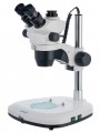
Stereoscopic. Microscopes designed to obtain volumetric images (usually in overhead, reflected light). The traditional design of such a microscope includes a pair of lenses, paired eyepieces (binoculars), a relatively low magnification, a wide field of view, and a significant working distance (the distance from the lens to the object in question). Such features allow the user to observe a three-dimensional image with a good depth of field, and also, without any special obstacles, operate various tool
...s in the field of view and clearly control their movements. This is indispensable for tasks such as repairing watches and other mechanisms, soldering microcircuits, creating miniatures, etc.
Note that this category may also include devices of a simpler design, with only one eyepiece and lens. Such a microscope is considered stereoscopic if it has a wide field of view, low magnification and a large depth of field; In terms of image quality, it will inevitably be inferior to models with two lenses, but will still provide a certain impression of three-dimensionality. Of course, such “pseudo-stereoscopes” are not suitable for serious tasks such as jewelry or watchmaking - most of them are intended for children and are designed so that a young researcher can examine in detail a pebble, flower or other object that is not shaped preparation for a biological microscope.Magnification
The range of magnifications provided by the device is from minimum to maximum.
The magnification of the microscope is calculated by the formula "the magnification of the eyepiece is multiplied by the magnification of the objective." For example, a 20x objective with a
10x eyepiece will give a magnification of 10*20 = 200x. Modern microscopes can be equipped with multi-objective turrets, zoom lenses (see below) and interchangeable eyepieces — so that in most models the magnification can be adjusted. This allows you to adjust the device to different situations: when you need to consider small details, a high degree of magnification is used, but to expand the field of view, the magnification must be reduced.
Detailed recommendations on optimal multiplicities for different tasks can be found in special sources. Here we note that many manufacturers go to the trick and indicate the maximum value of the magnification by the degree of magnification achieved with an additional Barlow lens. Such a lens can indeed give a serious increase in magnification, but it is not a fact that the image will turn out to be of high quality; for more details, see "Complete set".
Research method
Research methods applicable to this microscope model.
— Bright field. The most famous and widely used method of light microscopy. The object under consideration in such studies is placed on a light background, against which it looks darker. Note that different methods of illumination can be used for research: direct through, oblique, reflected. The first option (when the light from a lamp or a mirror under the stage shines through the sample) is optimal for studying transparent samples, the key details of which are darker than the general background. Typical examples are thin sections of animal and plant tissues. Oblique light is similar in application specifics, while it gives a grey background and is inferior to direct light in terms of backlight efficiency, but provides a more embossed image. As for reflected light, in this case it is indispensable when examining opaque objects: samples of ores and other materials, semiconductor wafers, etc. Anyway, bright-field microscopy well reveals, first of all, details that are noticeably different in light transmission or refractive index from the surrounding background (with through illumination), or give noticeable reflections / shadows (with reflected light).
— Dark field. A kind of opposite of bright-field research: the object under consideration or its individual elements are lighter than the surrounding background. However, this is not just a “negative” of the image, but a separate method with its own c...haracteristics. Illumination in dark-field microscopy is usually through, but it is carried out in a specific way: the middle of the light beam is blocked by a hood, and the light “cylinder”, passing through the condenser lens, turns into an “hourglass”. At the same time, in the narrowest place of such a "clock" there is a preparation, and towards the lens, the light cone expands so that it does not fall into the optics. Thus, the user sees in the microscope only the light scattered by the preparation and the dark background around. This method of research, among other things, allows you to identify "smooth" details that do not stand out sharply against the surrounding background and are not visible in a bright-field study. Among the applications of dark-field microscopy are the work with unstained biological preparations (cells, tissue samples, microorganisms), as well as the study of some transparent materials for small surface defects.
— Phase contrast. A method used to study transparent and colourless objects with an inhomogeneous structure, used when this inhomogeneity cannot be detected by more traditional bright-field microscopy. The idea of this method is that when passing through structures with different refractive indices, light receives different phase changes. These changes are not visible with ordinary optics, but they can be made visible with the help of special equipment — namely, a condenser and a lens of a special design. Accordingly, such equipment is necessarily included in the scope of delivery of the microscope.
— Fluorescent. This method provides the illumination of observed objects with light of a certain wavelength, under the influence of which these objects or their individual elements begin to glow, while the background remains dark. If necessary, coloring substances are introduced into the preparation to improve luminosity (a typical example is biological objects, most of which fluoresce rather weakly by themselves). For illumination, usually, UV radiation is used, therefore this method is also called ultraviolet microscopy; The image enters the eyepiece of the microscope through a filter that filters out UV rays, but freely passes the visible glow of the drug.
One of the main features of fluorescence microscopy is high resolution: it allows you to clearly see even very small objects that are not visible in the usual visible range. In fact, this method in terms of resolution is between classical optical and electron microscopy; At the same time, in contrast to electron and atomic microscopes, devices with the support of the UV method make it possible to examine even the hardware of living cells and microorganisms. And some special variants of this technique make it possible to achieve not micro, but nanoscopic magnifications. The second popular use of fluorescence microscopy is the detection of particles, elements, inclusions, etc., which are not visible under ordinary light, but stand out well in ultraviolet light. A typical example is the surface of many metals and alloys.
Turret
The number of lenses in the microscope turret.
The turret is a round nozzle with several lenses of different magnification. By turning such a nozzle, you can change the lens currently used; and the more lenses, the wider the choice for the user when choosing the optimal magnification of the microscope. On the other hand, numerous optics affects the dimensions and price of the device. In light
of this, most modern microscopes have
3-4 lenses — this number is considered optimal in terms of functionality and price.
Eyepiece
—
Monocular. An eyepiece with a single lens that can only be viewed with one eye. For obvious reasons, it is only used in biological microscopes (see "Type"). The advantages of monoculars are primarily smaller size and cost than other varieties; in addition, they do not require adjustment for interpupillary distance. On the other hand, constantly looking into the eyepiece with one eye is tiring, so this option is poorly suited for situations where you have to look into the microscope often and for a long time.
—
Binocular. Dual eyepiece that can be viewed with both eyes at once. Note that such optics are used not only in stereomicroscopes, originally intended for viewing an object through two lenses (see "Type"), but also in biological microscopes with one lens. The fact is that looking into an optical device with two eyes is much more convenient than with one, while the eyes are less loaded and fatigue does not occur so quickly. Therefore, for serious tasks associated with frequent use of a microscope, binoculars (or trinoculars, see below) are the best option. Such optics cost more than monocular, but this is offset by ease of use.
—
Trinocular. A kind of binocular (see the relevant paragraph), supplemented by a third optical channel for a special camera-video eyepiece. Such a camera is usually connected to a PC or laptop; by installing it in the soc
...ket for the third eyepiece, you can take photos and videos, as well as display the image in real time on the computer screen. At the same time, you can look through the microscope in the usual way. Devices with trinoculars are very functional and versatile, but they are complex and expensive.
— LCD screen. The microscope has an LCD screen that replaces the traditional eyepiece. You do not need to bend over to such a device each time to view the image, which is very convenient if observations need to be combined with record keeping and other similar activities. Microscopes of this design usually have a photo and video function, as well as various built-in tools — for example, a scale grid for estimating the size of visible objects, displayed directly on the screen. In addition, the image on the screen can be seen not only by the direct user, but also by everyone who is nearby; such features are indispensable during training sessions, consultations, presentations, etc. On the other hand, such microscopes turn out to be bulky and expensive.
— magnification factor. The magnification provided by the eyepiece. This parameter, along with the lens magnification, affects the overall magnification of the device (see above). The classic option for eyepieces in microscopes is 10x, but higher values \u200b\u200bare also found. The package may include several eyepieces, of different magnification — to change the overall degree of magnification. There is a multiplicity designation with a letter index, for example, WF10x. This means that the eyepiece has an extended field of view (WF — wide, EWF — extra wide, UWF — extra wide).
— Eyepiece tilt. The tilt of the eyepiece determines the position of the observer's head when looking through the microscope and the overall usability. According to this indicator, three main options can be distinguished: fixed angle, adjustable angle, without tilt. The fixed angle is most often 30° or 45° relative to the horizontal, these values are considered the most convenient. In angle-adjustable microscopes, the entire stand, with tube and stage, is fixed to the base with a swivel mount. This is the most convenient option, allowing you to adjust the tilt to your preference, but the mount tends to become loose over time, so it is rarely used in professional microscopes. The third variety — vertical microscopes, without tilt — have not received much distribution: this design is used in some stereoscopic models (see "Type") in order to ensure that the stage remains strictly horizontal (this is important for some work with microscopic objects).
— Rim diameter. The nominal diameter of the eyepiece used in the microscope, as well as the diameter of the hole in the tube, designed to install the eyepiece. Several standard diameters are used in modern microscopes, in particular 23 and 27 mm. In fact, this parameter is necessary, first of all, if you plan to purchase spare or replacement eyepieces for the microscope, or if you already have an eyepiece on the farm, and you need to evaluate its compatibility with this model.
— Diopter adjustment. The range of diopter correction provided in the eyepiece. This correction is used so that a nearsighted or farsighted person can look through the microscope without glasses or contact lenses. In most models with this function, the correction range is about 5 diopters in both directions; this allows the microscope to be used for low to moderate myopia/farsightedness.Interpupillary distance
Interpupillary distance in a microscope equipped with an eyepiece "under two eyes" — a binocular or trinocular.
In fact, this paragraph indicates the distance between the optical centers of the eyepieces. For normal visibility, it must exactly match the distance between the pupils of the user's eyes — hence, among other things, the name "interpupillary". And since the distance between the pupils can vary markedly for different people, then in all modern microscopes (for which this is generally relevant), the eyepieces are made movable, and the width of their location can be adjusted. In this paragraph, respectively, the range of such adjustment is indicated. Most often, it ranges from 55 to 75 mm — this is quite enough to choose the option for almost any adult user. But there are also more extensive adjustment ranges, mainly with an extension to the smaller side — for example, 52 – 76 mm or 48 – 75 mm. Such characteristics may be useful, in particular, when it comes to a children's microscope.
Maximum working distance
The greatest working distance provided by the microscope.
The working distance is the distance from the lens to the object in question. This parameter is important primarily for stereomicroscopes (see "Type"): the more space remains under the lens, the more convenient it is to work with various tools and devices in the field of view of the device. However, here it should be taken into account that the maximum working distance is achieved at the minimum magnification factor, as the magnification increases, the lens has to be brought closer to the object in question. For biological microscopes, the working distance does not really matter: such devices work mainly with flat preparations, to which the lens can be brought almost close.
Object table
The design of the object stage provided in the microscope.
— Stationary. Subject table, fixed motionless; focus in such microscopes is carried out by moving up and down the tube with the objective and the eyepiece. Such systems are simple and inexpensive, but focus while looking through a constantly moving eyepiece is not very convenient. In addition, for advanced biological microscopes (see "Type") with binoculars and trinoculars (see "Eyepiece"), this option is also poorly suited for some design reasons. But the vast majority of stereomicroscopes are equipped with stationary tables — this is the most reasonable design, taking into account the specifics of the application.
—
Movable. In microscopes of this type, the entire optical system is fixedly fixed on a tripod, and the stage can be moved up and down to focus the optics. This design is found exclusively in biological microscopes (see "Type"). It is somewhat more complicated and expensive than with a fixed table, but at the same time it is much more convenient: when focus, the eyepiece does not move, which allows you to comfortably adjust the image without looking up. In addition, it is the movable stage that is most suitable for advanced devices with binoculars and trinoculars (see "Eyepiece"), almost all such microscopes have such equipment.