Comparison Levenhuk Rainbow 2L vs Levenhuk D50L Plus
Add to comparison | 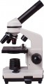 | 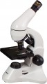 |
|---|---|---|
| Levenhuk Rainbow 2L | Levenhuk D50L Plus | |
from £76.50 | from £124.98 | |
| TOP sellers | ||
| Features | children's training | training |
| Type | biological | biological |
| Operation principle | optic | optical-digital |
| Magnification | 40 – 400 x | 64 – 1280 x |
| Research method | light field | light field |
Lens and eyepiece | ||
| Turret | 3 lenses | 3 lenses |
| Lens | 4x, 10x, 40x | 4x, 10x, 40x(s) |
| Eyepiece | monocular WF10x 45° incline diameter 23.2 mm | monocular WF16x 45° incline diameter 23.2 mm |
| Rotary eyepiece | ||
Design | ||
| Object table | mobile 90x90 mm | mobile 90x90 mm |
| Focus | coarse | coarse |
| Backlight | lED | lED |
| Top illumination | ||
| Bottom illumination | ||
| Condenser | N.A.=0.65 | N.A.=0.65 |
| Diaphragm | flat | flat |
| Features | brightness control | brightness control photo/video recording |
| Connection interfaces | USB | |
General | ||
| Power source | mains 230 V batteries | mains 230 V USB port batteries |
| In box | accessories and preparations set | camera accessories and preparations set Barlow lens cover/case |
| Material | plastic | metal |
| Added to E-Catalog | september 2017 | september 2017 |
Compare Levenhuk Rainbow 2L and D50L Plus
You may be interested in
My comparisons
Levenhuk Rainbow 2L often compared


Levenhuk D50L Plus often compared

Glossary
Features
General purpose of a microscope.
Nowadays, there are 4 main options for the appointment: children's, educational, laboratory and specialized microscopes. At the same time, different options (at least from the first three) may well be combined in one model — for example, the simplest and most inexpensive educational microscopes may well be positioned as children's, and the most advanced as laboratory ones. Here is a detailed description of the different destinations:
— Children's. The most simple and inexpensive microscopes, designed primarily for children who are taking their first steps in the natural sciences (as well as for other undemanding users who do not need particularly advanced functionality). Accordingly, such devices lack advanced features such as focus lock, Keller lighting, video outputs (for digital and opto-digital models), a trinocular with the ability to connect a camera, etc. In addition, the body can be made in bright colours, and in plastic is usually used as the body material. However, many children's microscopes are equipped with turrets for quick re-tuning of magnification, and the total magnification factor may well exceed 600x out of the box and 1000x in the top configuration.
— Educational. Microscopes well suited for teaching applications; sometimes such an appointment is even di...rectly indicated by the manufacturer. The specific functionality of such models is quite diverse, the type can also be different (both biological and stereoscopic). In general, devices of this specialization occupy an intermediate position between simple and inexpensive children's microscopes and advanced laboratory equipment. At the same time, there are many models that have a combined purpose — "children's / educational" or "training / laboratory". The first variety is simple and inexpensive, for educational purposes it is suitable mainly for school; the second option, in turn, can be useful even at the university faculty of natural sciences.
— Laboratory. The most advanced type of modern microscopes, designed for full-fledged laboratory research and other serious tasks. Accordingly, such models are not cheap, but they provide a high-quality image and, in general, have the most extensive functionality (although the specific set of features, of course, may be different). Among the features found in laboratory microscopes are a movable stage, installation of light filters, 2 types of illumination (lower and upper), Keller illumination, suitability for special microscopy methods (fluorescence, phase contrast), etc.
— Specialized. Microscopes of a specific design and purpose, one way or another different from more traditional models. These differences may vary; accordingly, the specific specialization also differs. So, recently, portable models for smartphones have gained quite significant popularity: with the help of a special clothespin, such a device is attached directly to the gadget opposite the main camera, and the smartphone screen plays the role of an eyepiece. Another popular variety is compact digital microscopes without their own screens, connected to PCs or laptops via USB, and even to smartphones via Wi-Fi (including via the Internet). This also includes professional equipment with a fairly narrow specialization: stereoscopes with special mounts for dental prosthetics, for soldering microcircuits, etc.; microscopes for metallurgical research; devices on a tripod with a remote rod, designed to inspect individual areas on general objects; comparative microscopes for ballistic and trace investigations in forensics; and etc.
Operation principle
— Optical. Traditional microscopes, which work based on the use of lenses and other optical elements. They allow you to provide high image quality and a good magnification factor, while they do not depend on electricity (except that batteries may be needed for the backlight system). This type of microscope uses traditional eyepieces, but there are some models that allow you to connect an external camera and display the image on a computer display. Also note that this is the only principle used in stereoscopic models (see "Type")
— Digital. Microscopes of this type are actually digital cameras, supplemented with powerful magnifying optics. The image from such a camera must be displayed on the screen; some models have their own displays, others do not have displays and need to be connected to a computer/laptop. The advantage of the first variety is independence from external equipment, the advantages of the second option are compactness and relatively low cost. At the same time, it should be noted that in terms of magnification, most digital microscopes are inferior to optical ones, and this principle is not suitable for a stereoscopic image.
— Optical-digital. Microscopes that combine the features of optical and digital models (see relevant paragraphs). Such models differ from "purely digital" devices by more advanced optics, with a revolving head and a...high magnification; from optical ones — with a built-in camera and using a screen as an eyepiece (traditional eyepieces are not used in optical-digital models).
Magnification
The range of magnifications provided by the device is from minimum to maximum.
The magnification of the microscope is calculated by the formula "the magnification of the eyepiece is multiplied by the magnification of the objective." For example, a 20x objective with a 10x eyepiece will give a magnification of 10*20 = 200x. Modern microscopes can be equipped with multi-objective turrets, zoom lenses (see below) and interchangeable eyepieces — so that in most models the magnification can be adjusted. This allows you to adjust the device to different situations: when you need to consider small details, a high degree of magnification is used, but to expand the field of view, the magnification must be reduced.
Detailed recommendations on optimal multiplicities for different tasks can be found in special sources. Here we note that many manufacturers go to the trick and indicate the maximum value of the magnification by the degree of magnification achieved with an additional Barlow lens. Such a lens can indeed give a serious increase in magnification, but it is not a fact that the image will turn out to be of high quality; for more details, see "Complete set".
Lens
— Zoom lens. Lens with variable magnification. Such optics allow you to smoothly change the overall magnification of the microscope within certain limits, without changing the objective/eyepiece and without even looking up from observations. On the other hand, zoom lenses are more complicated and more expensive than constant magnification optics. Therefore, they are mainly used in stereoscopic microscopes (see "Type"): in the repair, assembly and other tasks for which such devices are used, the ability to smoothly adjust the multiplicity is extremely useful.
— magnification factor. The magnification provided by the lens. This parameter, along with the magnification of the eyepiece, affects the overall magnification of the device (see above). Most biological microscopes (see "Type") are equipped with several different magnification objectives on the turret; this allows you to adjust the degree of magnification as desired by the user. The standard magnification options for such lenses are 4x, 10x, 40x, 100x.
— Achromat. One of the varieties of colour correction used in lenses. The need for colour correction is due to the fact that light of different colours is refracted differently by lenses, and without additional measures, the image in the microscope would be blurred with iridescent stains. Achromatic is one of the simplest types of colour correction; in such optics, colour distortions in yellow and green are corrected.... Achromatic lenses have simple design and low cost. However the image quality in them is far from perfect: such a lens gives a clear image only in the centre of the image, the width of the sharpness zone is about a third of the total width of the field of view, and red-blue streaks may appear along the edges of the image. However, this is quite enough for general acquaintance, initial training, and often for more serious tasks.
— Planachromat. An improved and improved version of achromatic lenses (see above). Plan achromats provide additional correction of the field curvature, due to which the area of a clearly visible image in such lenses is at least 2/3 of the total width of the field of view, and often even more. It is these lenses that are recommended for serious study and professional use.
— Rim diameter. The size of the thread used to mount the lens. A larger bore usually means a wider lens, which means higher aperture and better image quality. On the other hand, the large size affects the dimensions, weight and cost of optics. In modern microscopes, diameters from 20 to 35 mm are mainly found. Knowing the size of the thread, you can purchase replacement or spare lenses for the device.
Eyepiece
— Monocular. An eyepiece with a single lens that can only be viewed with one eye. For obvious reasons, it is only used in biological microscopes (see "Type"). The advantages of monoculars are primarily smaller size and cost than other varieties; in addition, they do not require adjustment for interpupillary distance. On the other hand, constantly looking into the eyepiece with one eye is tiring, so this option is poorly suited for situations where you have to look into the microscope often and for a long time.
— Binocular. Dual eyepiece that can be viewed with both eyes at once. Note that such optics are used not only in stereomicroscopes, originally intended for viewing an object through two lenses (see "Type"), but also in biological microscopes with one lens. The fact is that looking into an optical device with two eyes is much more convenient than with one, while the eyes are less loaded and fatigue does not occur so quickly. Therefore, for serious tasks associated with frequent use of a microscope, binoculars (or trinoculars, see below) are the best option. Such optics cost more than monocular, but this is offset by ease of use.
— Trinocular. A kind of binocular (see the relevant paragraph), supplemented by a third optical channel for a special camera-video eyepiece. Such a camera is usually connected to a PC or laptop; by installing it in the soc...ket for the third eyepiece, you can take photos and videos, as well as display the image in real time on the computer screen. At the same time, you can look through the microscope in the usual way. Devices with trinoculars are very functional and versatile, but they are complex and expensive.
— LCD screen. The microscope has an LCD screen that replaces the traditional eyepiece. You do not need to bend over to such a device each time to view the image, which is very convenient if observations need to be combined with record keeping and other similar activities. Microscopes of this design usually have a photo and video function, as well as various built-in tools — for example, a scale grid for estimating the size of visible objects, displayed directly on the screen. In addition, the image on the screen can be seen not only by the direct user, but also by everyone who is nearby; such features are indispensable during training sessions, consultations, presentations, etc. On the other hand, such microscopes turn out to be bulky and expensive.
— magnification factor. The magnification provided by the eyepiece. This parameter, along with the lens magnification, affects the overall magnification of the device (see above). The classic option for eyepieces in microscopes is 10x, but higher values \u200b\u200bare also found. The package may include several eyepieces, of different magnification — to change the overall degree of magnification. There is a multiplicity designation with a letter index, for example, WF10x. This means that the eyepiece has an extended field of view (WF — wide, EWF — extra wide, UWF — extra wide).
— Eyepiece tilt. The tilt of the eyepiece determines the position of the observer's head when looking through the microscope and the overall usability. According to this indicator, three main options can be distinguished: fixed angle, adjustable angle, without tilt. The fixed angle is most often 30° or 45° relative to the horizontal, these values are considered the most convenient. In angle-adjustable microscopes, the entire stand, with tube and stage, is fixed to the base with a swivel mount. This is the most convenient option, allowing you to adjust the tilt to your preference, but the mount tends to become loose over time, so it is rarely used in professional microscopes. The third variety — vertical microscopes, without tilt — have not received much distribution: this design is used in some stereoscopic models (see "Type") in order to ensure that the stage remains strictly horizontal (this is important for some work with microscopic objects).
— Rim diameter. The nominal diameter of the eyepiece used in the microscope, as well as the diameter of the hole in the tube, designed to install the eyepiece. Several standard diameters are used in modern microscopes, in particular 23 and 27 mm. In fact, this parameter is necessary, first of all, if you plan to purchase spare or replacement eyepieces for the microscope, or if you already have an eyepiece on the farm, and you need to evaluate its compatibility with this model.
— Diopter adjustment. The range of diopter correction provided in the eyepiece. This correction is used so that a nearsighted or farsighted person can look through the microscope without glasses or contact lenses. In most models with this function, the correction range is about 5 diopters in both directions; this allows the microscope to be used for low to moderate myopia/farsightedness.
Rotary eyepiece
This feature means that the eyepiece that the microscope is equipped with can rotate around a vertical axis — in other words, right and left. Typically, the range of rotation is a full 360°.
The swivel head of the eyepiece does not affect the main features and capabilities, but provides additional convenience for the user: the eyepiece can be rotated to the optimal position depending on the situation. This can be useful, for example, when two students or laboratory assistants sitting next to each other use one microscope with a preparation for two — if necessary, each can turn the eyepiece towards him without moving the entire device. The reverse side of this advantage is some complication of the design and an increase in its price.
The swivel head of the eyepiece does not affect the main features and capabilities, but provides additional convenience for the user: the eyepiece can be rotated to the optimal position depending on the situation. This can be useful, for example, when two students or laboratory assistants sitting next to each other use one microscope with a preparation for two — if necessary, each can turn the eyepiece towards him without moving the entire device. The reverse side of this advantage is some complication of the design and an increase in its price.
Bottom illumination
Bottom lighting is a lighting system, the light from which is directed from the bottom up.
In conventional (not inverted) microscopes, such illumination is directed towards the objective through a hole in the stage. It is this type of illumination that is used for classical bright-field microscopy using through illumination; Thus, the lower arrangement of the illumination is traditional for biological microscopes and is provided for in most of these models. But the presence of this function in "stereoscopes" is not typical, although it also occurs.
In turn, in inverted microscopes, the upper and lower illuminations are actually “swapped”. Accordingly, in such models, this function is intended for examining preparations (mostly opaque) in reflected light, and the light flux is directed from the lens to the preparation.
Features
— Adjustment of interpupillary distance. The ability to change the distance between the eyepieces in a binocular or trinocular microscope (see "Eyepiece"). For normal visibility, it is necessary that the distance between the lenses of the eyepieces correspond to the distance between the pupils of the user. This distance varies from person to person, so this setting may be required for comfortable use.
— Brightness adjustment. The ability to change the brightness of the backlight — to adjust the lighting to the specifics of the situation. For example, to study a thin transparent preparation in a bright field, high brightness will be unnecessary, but when transilluminating a dense dark object, it is indispensable.
— Illumination according to Keller. The presence of illumination in the microscope according to the Keller system. Such lighting is used exclusively in biological models (see "Type"), it is a sign of a professional level device. The Keller system complicates and increases the cost of the design, in addition, it may require specific settings, but with the right settings, the quality of the lighting is very high, and the image is as reliable as possible. Note that in microscopes there is a so-called. "simplified Keller system", when the settings are set at the factory and cannot be changed; however, in this case, it is precisely the full-fledged,...adjustable Keller lighting that is meant.
— Photo / video recording. Possibility of photo and video filming of the image visible in a microscope. Features of the implementation of this function in different microscopes may be different. For example, some models need to be connected to a computer, while others can record materials directly to a memory card or other media. Also, the cameras themselves, carrying out the shooting, can be both built-in and removable (see "Packaging" / relevant paragraphs).
Connection interfaces
Ways to transfer data to other devices provided in the design of the microscope.
This parameter is relevant primarily for digital and opto-digital models, as well as for individual optical devices equipped with cameras. All described microscopes can be equipped with AV and HDMI outputs, universal USB ports, removable media card readers, and Wi-Fi wireless modules. Here is a detailed description of each interface:
— AV output. Analogue output for video signal transmission. It is used primarily for live transmission of images from a microscope camera, and in some models — also for viewing footage stored in memory. Such outputs do not support HD resolutions and, in general, are inferior to HDMI in terms of overall “picture” quality (with the same camera characteristics). On the other hand, specifically for microscopes, these moments are not so often critical; analogue connectors are still quite popular in both conventional video equipment and special equipment; and the implementation of this interface is inexpensive. Therefore, AV outputs can be found even in fairly advanced models.
— HDMI. Digital output for video signal transmission. Similarly, AV can be used both for real-time broadcasting and for using the microscope as a video player when viewing saved materials (if such...a possibility is provided for in this model at all). At the same time, such outputs are more advanced than analogue AV: HD-quality images (including Full HD and higher) can be transmitted via HDMI, and the signal is very resistant to interference. We also recall that this interface is extremely common in modern video technology — in particular, the presence of at least one HDMI input is almost mandatory for TVs and monitors that support HD standards. On the other hand, the implementation of HDMI is noticeably more expensive, and it makes sense to use it with fairly advanced cameras, which in themselves significantly affect the price of microscopes. Therefore, such outputs can be found mainly in rather expensive and advanced devices.
— USB. Universal connector that allows different applications; a specific set of these options is directly related to the functionality of the microscope. Typical examples of using USB include the following: copying captured photos and videos to a computer or laptop; live image broadcast; remote control via PC / laptop (for example, moving the parent drug); charging the built-in battery, etc. The specific type of USB connector in the microscope may vary, however, usually, an appropriate cable is supplied in the kit for connecting to a standard full-size port.
— Card reader. The device for working with memory cards is usually SD, and in miniature pocket models — microSD. Such cards usually contain materials captured by the camera. In general, this function makes it much easier to copy information to other devices that also have card readers — primarily laptops and PCs; and miniature microSD cards are also supported by smartphones, tablets and other portable gadgets. Anyway, removing the card from the microscope and installing it in another device is often easier and faster than fiddling with a wired or Wi-Fi connection.
— Wi-Fi. A wireless module, which in this case is mainly used to communicate with an external device — such as a smartphone, laptop or PC. A Wi-Fi connection allows you to at least broadcast the image from the camera and copy the photos taken by it, and often also control other functions and settings (light brightness, movement of the driver, etc.). At the same time, the absence of wires provides additional freedom of movement and overall convenience. However, note that the specific communication format may be different, it should be specified separately. So, some models support only direct connection over a relatively short distance (in fact, up to a couple of tens of metres, or even less). Others are able to connect to an external device via the Internet, and here the distance does not play a role — there would be access to the World Wide Web. Still others allow both formats of work. Also note that individual devices with this function do not have their own screens at all and are designed for use with external gadgets; This design makes the microscope as compact and easy to carry as possible.